IgG selectivity
Assessing IgG Selectivity
There are several ways to establish the practical selectivity of anti-hapten IgGs. Competitive inhibition of binding is one of the simplest and can be performed by most users with high repeatability.
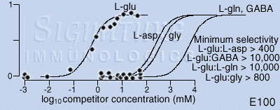
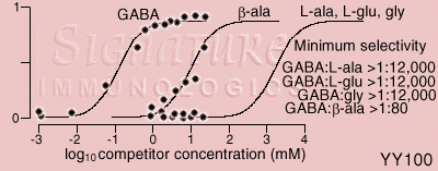
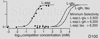
Method: Serial 250 nm sections of a tissue sample containing high levels of the target hapten are mounted on a 12-well slide probed with a 25 microliters of Signature Immunologics IgG in each well. A known concentration of free hapten (e.g. glutamate, GABA, etc.) is added to each well in 5 microliters. The slides are processed via standard HPI methods, and a constant target region (i.e. the same structure serially sectioned) captured with a CCD camera under radiometrically calibrated, constant gain, constant gamma conditions. The fractional inhibition (0=no detectable inhibition; 1=complete inhibition) is calculated and plotted on a semi-logarithmic scale. The minimum selectivity is calculated as the displacements of Langmuire adsorption isotherms for the target hapten and other suspected competitors.
Interpretation: The positioning of the adsorption isotherm for haptens showing no detectable inhibition is selected for the worst case selectivity. For example, the last aspartate point on the glutamate inhibition curve rises slightly. One cannot tell whether this is noise or a true deviation from baseline, but it is best to be conservative and position the rising slope of the function there. This leads to a minimum Glu:Asp selectivity of 400, but it could be much greater. This means that aspartate levels would have to exceed glutamate content by at least 400 fold to yield the same signal strength as glutamate. Many cells contain about 1 mM glutamate. Aspartate content would have to exceed 40 mM (a physiologically untenable level) to lead to a 10% signal corruption. Alternatively, the last GABA point on the glutamate inhibition curve indicates a minimum selectivity of >10,000, but again it could be much greater. In this case, GABA levels would have to reach 100 mM to generate but a 1% corruption in a 1 mM glutamate detection signal. For all practical biological purposes, these selectivities are virtually absolute.

© 2000 Signature Immunologics, Inc.
Copying and distribution:
This Technical Note may be copied and distributed for educational and non-profit
purposes as long as acknowledgement of Signature Immunologics' copyright is included
in every instance of use. It may not be sold or distributed for commercial gain.